Biological context
The cerebellar cortex is a subregion of the cerebellum structured as a folded sheet. This cortical sheet is itself decomposed into 2 major layers: the granular layer and the molecular layer, separated by a thin sheet of Purkinje cells, representing the Purkinje layer. Each layer contains its own cell types composition, placement rules and connectivity strategies. Fig. 1 shows the different cell type present in the network and their connections.
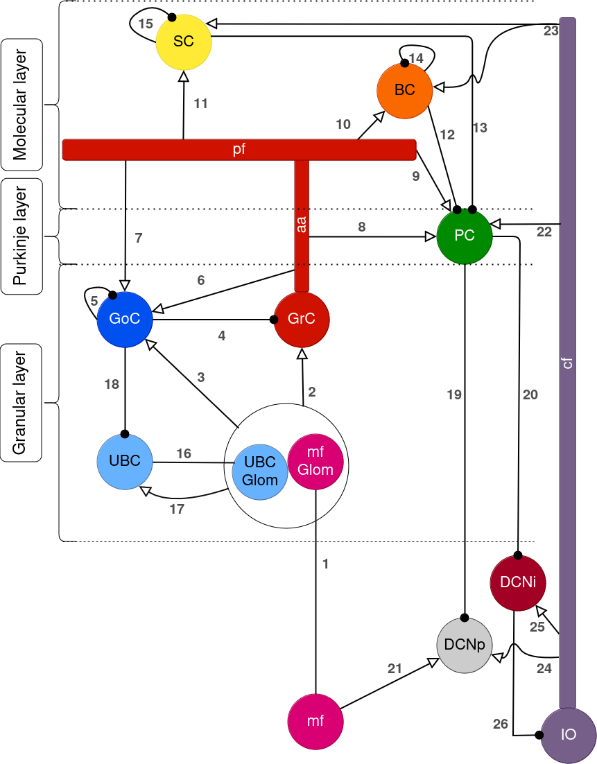
Fig. 1 Schematic representation of the cerebellar cortex neural network
Abbreviations: mf (Mossy fibers); glom (Glomeruli); UBC (Unipolar brush cells); GrC (Granule cells); GoC (Golgi cells); PC (Purkinje cells); aa (Ascending axons); pf (Parallel fibers); BC (Basket cells); SC (Stellate cells); IO (Inferior olive); cf (Climbing fibers); DCN (Deep cerebellar nuclei).
For most of the configurations available here, only a portion of the cerebellar cortex is built in a cubic volume with fixed layer thicknesses, to simplify the orientation and scaling of neuronal geometrical representation (morphologies) but also to limit the number of neurons and therefore the final size of the network.
Keep in mind that the default configurations do not include every cell type (and therefore connections) displayed in this Figure.
You can now head over to the Contents section.